Case Study: Dermoid Cyst Removal
Introduction
A dermoid cyst of the ovary is an abnormal growth that develops from egg cells and can contain various types of tissue, such as hair, skin, and teeth. Management of dermoid cysts usually involves surgical removal, either through an open or laparoscopic procedure. Medical management is ineffective in controlling the size of the cyst. If the cyst is small and not causing any symptoms, it may be monitored for a period of time to see if it resolves on its own. If the cyst does not resolve, or if it grows over time, surgical removal is usually recommended.
Case Report
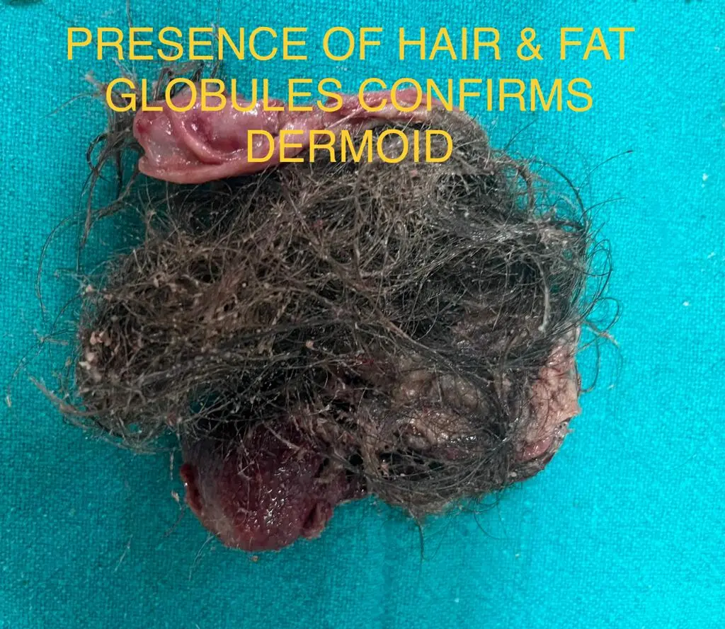
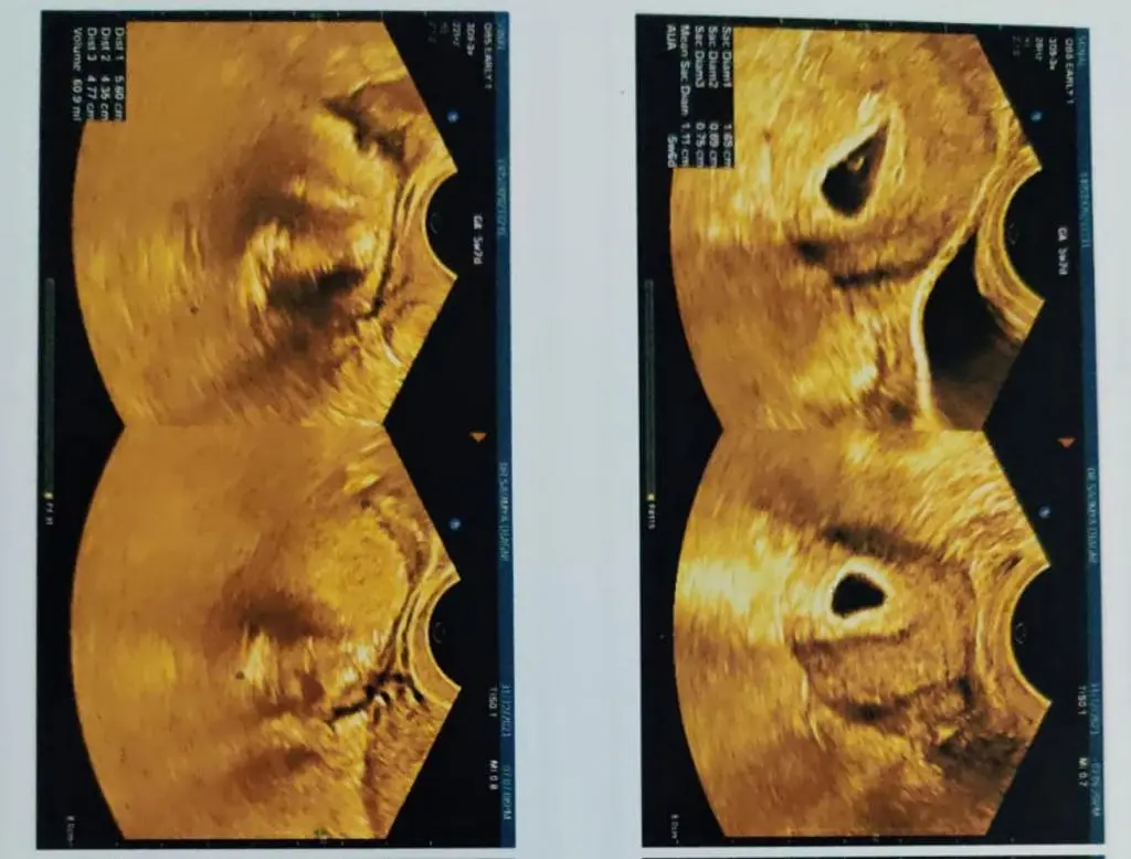
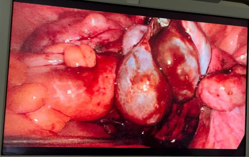
This patient who was detected with dermoid cyst first time during her pregnancy. It was an incidental finding and not giving any symptoms till then. Presence of fat globules and hair on ultrasound confirmed presence of dermoid. It enlarged mildly during pregnancy period and patient had 2-3 episodes of pain abdomen which subsided with pain killers, so surgery for its removal was not planned . She delivered normally at term. 5 months post delivery, she was taken up for laparoscopic cystectomy surgery for dermoid cyst removal. Cholecystectomy was also planned concurrently as pt had multiple gall bladder stones also. Post operatively, pt was fine and had uneventful recovery.


Conclusion
Dermoid cysts are common benign tumors that can occur in various parts of the body, including the ovaries. Surgical removal of the cyst may be necessary if it causes symptoms or if there is concern for malignancy. Laparoscopic cystectomy is a safe and effective option for the management of ovarian dermoid cysts, with excellent postoperative outcomes.

Case Study: Endometriosis
Case Report
A 38 year old lady, African black by ethnicity, had history of off and on abdominal pain for 6 months. Her family was complete with 3 kids. In initial work up- she was found to have bilateral complex masses in ovaries. She was referred from her country in view of raised CA 125 ( value 148) and fearing ovarian cancer.


Conclusion
In MRI pelvis here, masses were found to be blood filled and likely Endometriotic. She was posted for laparoscopic cystectomy for Endometriotic cysts. Per op , she had extensive adhesions between bowel and ovarian mass . After adhesiolysis, bilateral ovarian cystectomy done and post operatively GnRH injection given for further suppression. She is free of pain now and has been put on medical treatment for endometriosis suppression and sent back to her country.
Case Study: Multiple Uterine Fibroids
Case Report
In this section, we will discuss a case of multiple fibroids. A 30 year old female presented with heavy menstrual bleeding. MRI revealed multiple uterine fibroids. During work up , she was found to have very low Hb of 5 gm. Multiple blood transfusions were given . She was prepared for myomectomy or fibroid removal surgery. 16 fibroids of variable size ranging from 8 cm to 1 cm were extracted. Above case does clearly show need for more awareness about fibroids and how severe bleeding can affect our quality of life. Fibroids in the uterus, or uterine fibroids, are the benign, tumors in people of childbearing age. They are also known as leiomyomas and myomas.
Symptoms
Many people have fibroids with no symptoms, whereas others experience pain, bleeding, or both The symptoms of uterine fibroids can include:
- • heavy periods, also known as menorrhagia, which can lead to anemia
- • painful periods
- • lower backache or leg pain
- • constipation
- • discomfort or a feeling of fullness in the lower abdomen, especially in the case of large fibroids
- • frequent urination
- • pain during sexual activity, also known as dyspareunia
Some people may have fertility problems associated with fibroids. It is important to timely reach out to your doctor and get yourself treated. Dr Deepti Asthana is a senior gynaecologist and expert in laparoscopic as well as open surgery. Do visit her if you want genuine advise for your problem

Case Study: Abdominal Mass
Case Report
This is a case about a young married girl from pacific island who had a large abdominal mass from pelvis to epigastrium. It was a clear cystic mass with no signs of malignancy. Laparotomy revealed straw coloured fluid filled cystic mass arising from adenexa with multiple nodular deposits involving almost each and every organ of abdomen. Series of pathological tests revealed it to be tubercular. Patient started on ATT and went back to her country happily.

Large mass after removal of straw coloured fluid
